![]()
Vision:
An In-Depth Look at Eagle Eyes
Part 2
| What Lies Beneath | |||||
This diagram of an eagle eye and a human eye shows them as cross-sections, as if looking down on them from above the head. Look at your own eye in a mirror or look at one of your classmate's eyes. |
|||||
|
Try this!
|
|||||
| Iris: An Open and Shut Case | |||||
| Your
iris and a Bald Eagle's iris may be different colors, but they have
the same job: to control the amount of light that shines onto your
retina. There are two kinds of muscles in the iris. Circular muscles
encircle the iris close to the pupil, and straight muscles radiate
out like rays of the sun. When the inner, circular ones contract,
the iris gets bigger, making the pupil smaller. When the outer, radiating
ones contract, the iris gets smaller, making the pupil bigger. |
|||||
| Activity Work with a partner. Take turns watching your partner's pupil and iris change as the amount of light changes.
|
 |
||||
Cornea:
Window to the World When light passes through any curved lens, it bends. The bending of light through a convex lens like the cornea makes it "converge." The image formed by the cornea is upside down and reversed from right to left. |
|||||
| Lens:
Making Accommodations If the cornea were the only curved "window" that light passed through in the eye, far objects would focus very easily, but near objects would not. A human's cornea can't change its shape in order to bring objects into focus, but fortunately, one part of our eye CAN change shape. In order to help us focus on close objects, the LENS of our eye changes shape. This is called accommodation. Tiny fibers called ligaments and muscles change the shape of the lens, making it thinner to focus on far objects or thicker to focus on near objects. Eagles can change the shape of their lens, and can also change the shape of their corneas. This allows them more precise focusing and accommodation than we humans can get. |
|||||
Retina:
Where Vision Happens
|
|||||
Try
this!
Rod cells don't see color; they simply see light. And several rod cells network with each other, sending the brain messages on a single nerve. So vision with the rod cells isn't as precise, but is very fast. Rod cells may see only black and white, but they are extremely sensitive to light, so they help us see in the dark and notice quick movements. Eagles have a higher percentage of cone cells than we humans do, so they can't see as well as us at night, even if they do see better in daylight. If a human eye is shaped exactly right, things focus precisely on the retina. Sometimes the eye is longer than it should be, and the picture focuses in front of the retina. This condition is called "myopia" or nearsightedness. If the eye is shorter than it should be, the picture focuses behind the retina. This is called "hyperopia," or farsightedness. People wear glasses or contact lenses with exactly the right curve to move the focus onto the retina. Eagles with eyes that are shaped wrong can't wear glasses. Since good vision is so critical to their ability to get food, eagles with less than perfect vision quickly starve, and never get old enough to reproduce. So eagle parents all have great vision, and luckily their babies take after them! |
|||||
Fovea: Magnifying the View
|
|||||
The
Mysterious Pecten
Scientists have some data that supports the first four. The last two are simple guesses without evidence to support them. Which of these theories makes sense to you? |
|||||

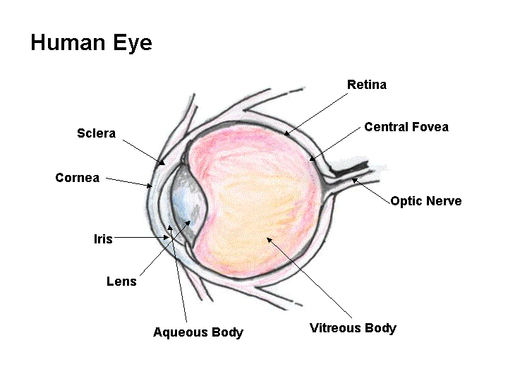


Discussion of Journaling Questions
- Click
on the diagram
of the human eye so you can see the large, labeled picture, and
compare it to your eye or a classmate's. Which of the labeled parts
can you actually see on a real eye? Which layers of the eye does light
pass through to reach the retina? Why does the pupil of a real eye look
so black?
Answer: What parts can we see on a real eye? If you look at a classmate's eye from the side, you might be able to see the clear cornea sticking out like a thin bubble. (You can't see your own cornea unless you use two mirrors, and even then it's almost impossible!) You can see the black pupil, which is really just the hole that lets light pass through the lens. You can see the colored iris. The lens is too clear to see at all. You can see the white sclera. The rest you just have to imagine!
- What eye
parts does light pass through? The cornea, aqueous body, lens, and vitreous
body.
Why does the eye look so black? Inside the sclera of the eye is a thin layer called the choroid coat, which has special pigments that make it look very dark. These pigments absorb extra light inside the eye so the only light we see is what is actually on the retina, giving us clearer vision.
- Which
muscles (the circular ones or the radiating ones) work when the light
suddenly gets brighter? Which work when the light suddenly gets dimmer?
Answer: When the light suddenly gets brighter, the circular muscles contract to close the pupil a bit. When the light suddenly gets dimmer, the radiating muscles contract to pull the pupil more open.
© 1997 – 2025 Journey North. All rights reserved.![]()
![]()